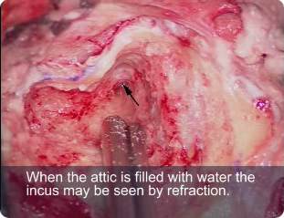
Step 5. Identify the incus (continued)
5.3 There will be another group between the sigmoid sinus and the ear canal. Depth removal of the cells may leave a groove formed by the facial nerve in front and sigmoid sinus behind. These cells may be continuous with cells that pass medial to the facial nerve then under the cochlea. Beware of the cortical bone over the jugular bulb in this region.
5.4 Display the medial aspect of the mastoid process and the digastric ridge.

|
