Step 5. Identify the incus
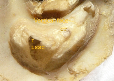
5.1 Prepare to expose the next landmark, the incus. It is exposed by removal of bone between the ear canal and floor of the middle fossa. It is accomplished with sweeps of the burr from inside outwards starting in the depth of the dissection and using the shoulder of the burr. The gap between the middle fossa and ear canal narrows laterally. The position of the incus is a guide to the depth of the facial nerve and semicircular canal.
5.2 Remove the air cells in the depth of the cavity. The semicircular canal region is on the medial side of the mastoid antrum. The antrum continues as a group of cells that extend back to the sinodural angle.
|