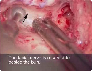
Step 6. Identify the facial nerve (continued)
6.6 During the dissection it is safe to expose the dura mater over the middle fossa and sigmoid sinus but it is not acceptable to expose the skin of the ear canal of the membranous part of the lateral canal. The facial nerve should only be exposed when the dissection is prepared for this step.
6.7 When the temporal bone is poorly pneumatised it is acceptable to expose the dura so the landmark gives the surgeon information about the depth of the next target feature.
6.8 If there are no retro-facial cells define the position of the facial nerve by displaying the plane of sigmoid sinus fascia. Having defined the fascia of the sigmoid sinus remove the bone anterior to it with movements parallel to it until the bone over the posterior and lateral aspect of the facial canal are displayed.

|
